
Knee - Anterior View
Ultrasound Images
Main Image

Annotations
Classification
Case code:
GN-ANT-M-A1016
Joint:
Knee
View:
Anterior
Severity:
Moderate
Patient
Age:
86 years
Sex:
Female
Condition:
Acquired hemophilia A (autoimmune, inhibitor positive)
Joint ROM:
15-115° (moderately reduced)


History
Elderly female patient diagnosed with acquired hemophilia A at age 82 following spontaneous extensive soft tissue hematomas and prolonged aPTT discovered during preoperative evaluation for cataract surgery. Inhibitor titer measured at 18 Bethesda Units. No previous personal or family history of bleeding disorders. Has experienced 4 episodes of spontaneous right knee hemarthrosis over the past 30 months despite immunosuppressive therapy (corticosteroids and cyclophosphamide) and bypassing agent coverage. Joint symptoms complicate pre-existing age-related osteoarthritis. Reports moderate pain (VAS 5-6/10) with daily activities. Uses walker for ambulation. Living independently with home care assistance three times weekly.
Physical Examination
Moderate knee swelling with palpable effusion. Age-related muscle atrophy of lower extremities with quadriceps showing additional wasting from hemophilic involvement. Mild valgus knee deformity. Range of motion limited by combination of hemarthrosis sequelae and degenerative changes: lacks 15° extension, achieves 115° flexion with discomfort. Crepitus present throughout range. Joint line tenderness medially and anteriorly. Gait significantly impaired requiring walker support with slow, cautious pattern. Difficulty with sit-to-stand transfers.
Ultrasound Findings
- Hoffa’s fat pad: moderate alterations combining hemophilic and degenerative changes
Anatomical Zones
Hoffa’s fat pad
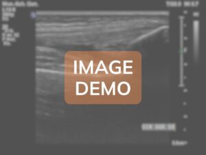
The adipose pad demonstrates moderate chronic alterations representing a complex interplay between acquired hemophilic arthropathy and age-related degenerative changes, with predominant hemosiderin deposition and fibrotic transformation.
Echostructure and Thickness:
Heterogeneous with moderate fibrosis; thickness 9.5mm (moderately thickened)
Findings:
The Hoffa's fat pad shows moderate pathological alterations with partial preservation of adipose tissue architecture. Approximately 50-60% of the fat pad volume maintains recognizable adipose echostructure (hyperechoic), while the remaining 40-50% demonstrates fibrotic transformation. The overall thickness measures 9.5mm, representing moderate enlargement compared to age-matched controls (expected 6-7mm in elderly patients). Multiple hypoechoic areas are distributed throughout the fat pad, with the largest measuring 12x7mm in the superior central portion, representing zones of fibrotic tissue replacement and hemosiderin deposition from recurrent hemorrhagic episodes. Several discrete hyperechoic foci without posterior shadowing are scattered within the fat pad, consistent with hemosiderin deposits rather than calcifications, appearing as 1-2mm punctate echogenic spots. Approximately 5-6 fibrous bands (thickness 1.5-2mm each) traverse the fat pad in various orientations, most prominently in the superior and lateral aspects, creating a reticular pattern that disrupts the normal homogeneous adipose appearance. These bands likely represent organized fibrous septa from previous hemorrhagic organization. The anterior border adjacent to the patellar ligament shows irregular interface with loss of the normal smooth plane, suggesting adhesion formation. The posterior border demonstrates moderate thickening and irregularity where it interfaces with the synovium. A small loculated fluid collection (5x3mm) is present in the lateral recess of the fat pad, possibly representing a small hemorrhagic cyst or loculated effusion. Color Doppler examination reveals moderate increase in vascularity with 4-5 small vessels (0.5-0.8mm diameter) penetrating into the fibrotic areas, indicating ongoing inflammatory activity despite the patient's age. The vascular pattern is less chaotic compared to younger patients with hemophilic arthropathy, possibly reflecting the slower disease tempo in acquired hemophilia. The fat pad demonstrates moderately reduced compressibility on probe pressure compared to expected in elderly patients, indicating significant fibrotic component. Dynamic examination during knee flexion reveals impaired mobility of the fat pad with restricted anterior displacement, contributing to anterior knee pain and impingement symptoms. The degree of fibrosis appears disproportionate for only 30 months of disease duration, suggesting that pre-existing age-related fat pad changes (fibrofatty involution commonly seen in elderly patients) may have been accelerated and exacerbated by the superimposed hemorrhagic episodes. Areas of the fat pad adjacent to the patellar ligament and tibial plateau show increased echogenicity and thickening, consistent with chronic mechanical stress and inflammation. No discrete calcifications or ossifications are identified, which is typical for the relatively short disease duration despite patient's age.
Direct Comparison
Compare this case with reference cases of different severity grades for in-depth analysis.
Cases available for comparison: 3 of 3 possible
Additional Notes
This case represents an uncommon presentation of acquired hemophilia A in an elderly female patient, with moderate hemophilic arthropathy developing in the context of pre-existing age-related joint changes. The score reflects moderate structural damage accumulated over relatively short disease duration (30 months), though the ultrasound findings show complex overlay of hemophilic changes upon baseline osteoarthritic alterations. The management is complicated by the patient's age, comorbidities (hypertension, type 2 diabetes, osteoporosis), and the challenges of immunosuppressive therapy in the elderly population. Current treatment includes prednisolone 20mg daily with gradual taper, rituximab infusions every 6 months for inhibitor eradication (current titer reduced to 4 BU), and recombinant activated factor VII (rFVIIa) for breakthrough bleeding episodes. The prognosis for complete inhibitor eradication is approximately 50-60% in acquired hemophilia, though elderly patients may have lower rates of immune tolerance. Joint preservation is a priority given limited surgical options at this age. Physical therapy has been adapted for elderly patient with focus on maintaining functional independence rather than athletic goals: seated strengthening exercises, assisted range of motion, and gait training with walker. Pain management includes scheduled acetaminophen 1000mg three times daily (avoiding NSAIDs due to bleeding risk), topical capsaicin cream, and occasional low-dose tramadol for severe pain episodes. Fall prevention strategies are critical given bleeding risk, including home safety assessment, proper lighting, removal of tripping hazards, and consideration of hip protectors. The patient's family has been educated about signs of acute hemorrhage requiring emergency evaluation. Quality of life considerations are paramount, with multidisciplinary team including hematology, geriatrics, physical therapy, and social work coordinating care. Follow-up ultrasound is planned in 6 months to assess progression, with particular attention to development of additional joint involvement. The case highlights the importance of maintaining high clinical suspicion for acquired hemophilia in elderly patients presenting with spontaneous bleeding, and the challenges of managing hemophilic arthropathy in this unique patient population where treatment goals must be balanced against age-related limitations and comorbidities.