
Ankle - Anterior View
Ultrasound Images
Main Image

Annotations
Classification
Case code:
CV-ANT-G-A1099
Joint:
Ankle
View:
Anterior
Severity:
Severe
Patient
Age:
46 years
Sex:
Male
Condition:
Hemophilia A severe (FVIII <1%), end-stage hemophilic arthropathy
Joint ROM:
Dorsiflexion 0°, Plantarflexion 25° (severely restricted)


History
Patient with severe hemophilia A diagnosed at 18 months following intracranial hemorrhage. Target joint development in right ankle began at age 7, with estimated 200+ hemarthrosis episodes throughout childhood and adolescence. Received on-demand treatment with cryoprecipitate until age 12, then plasma-derived concentrates. Prophylaxis initiated at age 18 but with poor adherence during young adulthood (age 18-28) due to psychosocial challenges. Compliance improved after age 30, but extensive joint damage already established. Reports severe chronic pain (VAS 8-9/10) requiring daily opioid analgesics. Previous interventions: radiosynovectomy at age 25, arthroscopic synovectomy at age 32 (temporary improvement). Currently on long-term disability, unable to work. Experiences 4-5 breakthrough bleeding episodes annually despite prophylaxis (FVIII 3000 IU three times weekly). Comorbidities include chronic pain syndrome, depression, hepatitis C (cured 2019), and hypertension.
Physical Examination
Severe ankle deformity with marked varus hindfoot alignment (15° varus). Massive swelling with tense effusion and prominent circumferential synovial thickening. Significant muscle atrophy of entire lower leg with calf circumference 6cm smaller than contralateral (25cm vs 31cm). Skin changes include hyperpigmentation and thinning. Fixed equinus contracture with ankle resting in 25° plantarflexion, unable to achieve neutral position. Range of motion severely restricted: complete loss of dorsiflexion (0°), plantarflexion limited to 25° from resting position (total arc only 25°). Rigid end-feel suggesting bony ankylosis and severe capsular contracture. Joint instability with medial-lateral laxity and positive anterior drawer. Severe tenderness throughout. Gait severely antalgic requiring bilateral forearm crutches. Cannot perform single-leg stance or any heel raises. Moderate pedal edema (2+) extending to mid-calf.
Ultrasound Findings
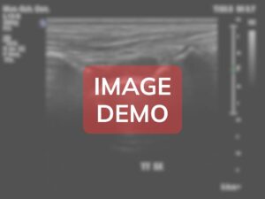
Ultrasound examination in anterior view reveals severe end-stage pathological changes:
- Anterior joint capsule: severe thickening (>8mm) with calcifications
- Anterior recess: massive synovial proliferation (>10mm) with complex organized effusion
- Talar dome: severe cartilage loss with near-complete destruction and exposed bone
- Distal tibial surface: extensive erosions, large osteophytes, subchondral cysts
- Marked synovial hyperemia with chaotic neovascularization
- Bony surfaces: extensive erosions, irregular contours, pseudocysts
- Large complex effusion with multiple loose bodies and debris
- Anterior soft tissue severe thickening and fibrosis
- Joint space collapse with areas of pseudoankylosis
Anatomical Zones
Tibiotalar joint anterior recess
The anterior recess demonstrates severe end-stage degenerative changes consistent with long-standing severe hemophilic arthropathy, showing massive synovial proliferation, near-complete articular cartilage destruction, extensive bony erosions, and structural disorganization representing decades of recurrent hemorrhagic insults.
Synovial proliferation and articular destruction:
11.5mm massive synovial proliferation; near-complete cartilage loss; extensive erosive changes
Findings:
The anterior recess displays catastrophic pathological alterations representing end-stage disease. The synovial membrane demonstrates massive proliferation measuring 11.5mm (normal 80% loss). Multiple large full-thickness defects (10+ areas ranging 5-15mm) expose severely irregular, eroded bone surfaces. The largest defect (15x8mm) creates deep excavation into subchondral bone. The talar subchondral bone shows catastrophic changes with multiple large erosions (8-12mm depth) creating "moth-eaten" appearance. Numerous subchondral pseudocysts (5-10mm diameter) represent bone necrosis and cystic degeneration. The distal tibial surface demonstrates similarly severe destruction with multiple erosions (5-12mm depth), near-complete cartilage loss (80% loss) with areas of apparent bone-on-bone contact suggesting pseudoankylosis. The anterior capsule demonstrates severe fibrotic thickening (8.5mm) with 15-20 calcific deposits (1-4mm) scattered throughout. Color Doppler reveals marked neovascularization with >15-20 vessels (0.5-2mm) creating dense chaotic network (grade 3-4 vascularity). Power Doppler shows confluent synovial blush. Spectral Doppler demonstrates low-resistance flow (RI 0.45-0.55) with elevated velocities (15-25 cm/sec) indicating active inflammation. Peri-articular soft tissues show severe thickening (8-10mm subcutaneous tissue) with obliterated tissue planes and widespread tenosynovitis in multiple extensor tendon sheaths.
Direct Comparison
Compare this case with reference cases of different severity grades for in-depth analysis.
Cases available for comparison: 3 of 3 possible
Additional Notes
This case represents end-stage hemophilic ankle arthropathy with score of 14/16 indicating near-maximal structural damage and profound disability. The extensive destruction reflects decades of inadequate hemostatic control, compounded by non-adherence during young adulthood. Current management focuses on palliation: maximal prophylaxis primarily prevents further bleeding rather than reversing damage. Pain management requires multimodal approach including long-acting opioids, gabapentin, duloxetine, and topical therapies, though control remains suboptimal (VAS 6-7/10). Ankle arthrodesis remains definitive surgical option but presents significant challenges: perioperative bleeding risk, poor bone quality with extensive erosions, severe soft tissue contracture, infection risk, and uncertain functional outcome. Patient has elected to defer surgery, preferring conservative management. Physical therapy focuses on maintenance goals: preserving residual motion, slowing atrophy, aquatic therapy, and gait training with bilateral crutches and custom AFO. Psychosocial support addresses severe depression and functional limitations through CBT, support groups, and vocational rehabilitation. Prognosis is poor for functional improvement without surgery, though even successful arthrodesis will leave permanent severe limitation. This case exemplifies devastating consequences of inadequately treated hemophilia and underscores the critical importance of early prophylaxis in preventing such catastrophic outcomes in modern hemophilia care.