
Clinical Cases Comparison
GN-ANT-L-A1012 - Mild
VS
GN-ANT-N-A0500 - Normal

Classification
Joint:
Knee
View:
Anterior
Severity:
Mild
Patient
Age:
24 years
Sex:
Female
Condition:
Moderate symptomatic hemophilia A carrier (FVIII 28%)
Annotations


Classification
Joint:
Knee
View:
Anterior
Severity:
Normal
Patient
Age:
36 years
Sex:
Male
Condition:
Healthy subject
Annotations
History
GN-ANT-L-A1012
Symptomatic carrier of hemophilia A with FVIII levels at 28%. History of menorrhagia since menarche treated with hormonal therapy. Presented 3 episodes of hemarthrosis in the left knee over the past 18 months, following minor trauma (fall during sports activity, direct contusion). Episodes were managed with RICE (rest, ice, compression, elevation) and single administration of FVIII concentrates. Reports occasional joint discomfort after prolonged physical activity. Has never followed regular prophylaxis.
GN-ANT-N-A0500
Healthy adult, office worker, engages in regular physical activity (running, gym) three times a week. No history of joint diseases, significant trauma, or knee surgery. Does not take any medication. Reports no pain, swelling, or functional limitations. Examination performed as a healthy control as part of a study aimed at defining reference ultrasound parameters in adults.
Physical Examination
GN-ANT-L-A1012
Knee appears normal on inspection without evident swelling. Mild tenderness on deep palpation of the suprapatellar region. No signs of acute joint effusion. Quadriceps muscle trophism preserved, symmetric compared to contralateral. Range of motion slightly reduced in maximum flexion (135° vs 140° expected) due to sensation of tension reported by patient. Ligamentous stability tests normal. Normal gait without limping. Mild discomfort during full squat.
GN-ANT-N-A0500
Knee with normal morphology on inspection, without signs of inflammation, deformity, or muscle atrophy. No joint effusion on palpation. Quadriceps trophism preserved. Full and painless range of motion. Ligament stability tests (Lachman, drawer, varus/valgus stress) negative. Normal gait without limping.
Ultrasound Findings
GN-ANT-L-A1012
- Quadriceps tendon: thickness at upper limit (7mm), mild focal echostructure heterogeneity
- Suprapatellar recess: mild synovial thickening (3mm), minimal fluid collection
- Patellar ligament: preserved morphology, mild localized thickening (5.5mm)
- Hoffa’s fat pad: preserved adipose echostructure with minimal focal fibrous bands
- Articular cartilage: preserved profile with minimal superficial irregularities
- Bony surfaces: regular, absence of significant erosions
- Minimal joint effusion (<3mm) in suprapatellar recess
- Mild increase in Doppler signal in synovium
GN-ANT-N-A0500
The ultrasound examination in the anterior projection shows:
- Quadriceps tendon: normal thickness and fibrillar echotexture
- Suprapatellar recess: empty, thin synovium (<1.5 mm)
- Patellar ligament: regular morphology, homogeneous echotexture
- Hoffa’s fat pad: normal adipose tissue without thickening
- Patellar cartilage: smooth and regular surface
- Femoral bone surfaces: sharp contours without irregularities. No effusion, intra-articular debris, or inflammatory signs
Anatomical Zones
🔄 Quadriceps tendon
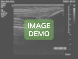
The tendon presents initial structural alterations compatible with previous stress from hemarthrosis, without significant functional compromise.
Findings: Mild tendon thickening with general preservation of normal parallel fibrillar echostructure. Presence of focal area of heterogeneity (8x3mm) in the lateral portion of the tendon, approximately 2cm proximal to patellar insertion, compatible with outcome of previous intratendinous edema/hematoma. Minimal scattered punctate calcifications (<1mm). Insertion on patellar base is regular without signs of enthesopathy. Doppler shows mild localized increase in vascularization in the heterogeneous area.

🔄

🔄
🔄 Quadriceps tendon
In this case, the quadriceps tendon shows normal ultrasound features typical of a healthy adult.
Findings: Well-defined parallel fibrillar echotexture. Homogeneous ultrasound pattern without areas of thickening or hypoechogenicity. Regular myotendinous junction. Clear and well-defined insertion at the base of the patella. No intratendinous calcifications or vascular alterations on Doppler.
🔄
Zone not present
ℹ️ Patellar ligament
The patellar ligament shows completely normal morphology and echotexture for the patient’s age.
Findings: Length of 48 mm with a uniform thickness of 4 mm. Straight course from the patellar apex to the anterior tibial tuberosity. Normal tendinous echotexture with clearly visible parallel fibers. No areas of degeneration, calcification, or discontinuity. Regular bony insertions without signs of enthesopathy. Normal vascularization on Doppler.
🔍
🔍

🔍
Zone not present
ℹ️ Hoffa’s fat pad
The infrapatellar fat pad shows the normal ultrasound features of mature adipose tissue.
Findings: Tessuto adiposo ipereoico omogeneo con normale morfologia triangolare. Contorni regolari e ben definiti. Assenza di ispessimenti, bande fibrotiche o aree di fibrosi. Non si evidenziano raccolte fluide o alterazioni strutturali. Vascolarizzazione fisiologica al Doppler senza segni di iperemia o neoangiogenesi. Rapporti normali con il legamento rotuleo e le strutture circostanti.
🔍
🔍
🔍
VS
Anatomical Zone
VS