
Clinical Cases Comparison
GN-ANT-M-A1016 - Moderate
VS
GN-ANT-N-A0500 - Normal

Classification
Joint:
Knee
View:
Anterior
Severity:
Moderate
Patient
Age:
86 years
Sex:
Female
Condition:
Acquired hemophilia A (autoimmune, inhibitor positive)
Annotations

Classification
Joint:
Knee
View:
Anterior
Severity:
Normal
Patient
Age:
36 years
Sex:
Male
Condition:
Healthy subject
Annotations
History
GN-ANT-M-A1016
Elderly female patient diagnosed with acquired hemophilia A at age 82 following spontaneous extensive soft tissue hematomas and prolonged aPTT discovered during preoperative evaluation for cataract surgery. Inhibitor titer measured at 18 Bethesda Units. No previous personal or family history of bleeding disorders. Has experienced 4 episodes of spontaneous right knee hemarthrosis over the past 30 months despite immunosuppressive therapy (corticosteroids and cyclophosphamide) and bypassing agent coverage. Joint symptoms complicate pre-existing age-related osteoarthritis. Reports moderate pain (VAS 5-6/10) with daily activities. Uses walker for ambulation. Living independently with home care assistance three times weekly.
GN-ANT-N-A0500
Healthy adult, office worker, engages in regular physical activity (running, gym) three times a week. No history of joint diseases, significant trauma, or knee surgery. Does not take any medication. Reports no pain, swelling, or functional limitations. Examination performed as a healthy control as part of a study aimed at defining reference ultrasound parameters in adults.
Physical Examination
GN-ANT-M-A1016
Moderate knee swelling with palpable effusion. Age-related muscle atrophy of lower extremities with quadriceps showing additional wasting from hemophilic involvement. Mild valgus knee deformity. Range of motion limited by combination of hemarthrosis sequelae and degenerative changes: lacks 15° extension, achieves 115° flexion with discomfort. Crepitus present throughout range. Joint line tenderness medially and anteriorly. Gait significantly impaired requiring walker support with slow, cautious pattern. Difficulty with sit-to-stand transfers.
GN-ANT-N-A0500
Knee with normal morphology on inspection, without signs of inflammation, deformity, or muscle atrophy. No joint effusion on palpation. Quadriceps trophism preserved. Full and painless range of motion. Ligament stability tests (Lachman, drawer, varus/valgus stress) negative. Normal gait without limping.
Ultrasound Findings
GN-ANT-M-A1016
- Hoffa’s fat pad: moderate alterations combining hemophilic and degenerative changes
GN-ANT-N-A0500
The ultrasound examination in the anterior projection shows:
- Quadriceps tendon: normal thickness and fibrillar echotexture
- Suprapatellar recess: empty, thin synovium (<1.5 mm)
- Patellar ligament: regular morphology, homogeneous echotexture
- Hoffa’s fat pad: normal adipose tissue without thickening
- Patellar cartilage: smooth and regular surface
- Femoral bone surfaces: sharp contours without irregularities. No effusion, intra-articular debris, or inflammatory signs
Anatomical Zones
🔄 Hoffa’s fat pad
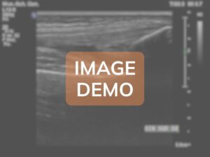
The adipose pad demonstrates moderate chronic alterations representing a complex interplay between acquired hemophilic arthropathy and age-related degenerative changes, with predominant hemosiderin deposition and fibrotic transformation.
Findings: The Hoffa's fat pad shows moderate pathological alterations with partial preservation of adipose tissue architecture. Approximately 50-60% of the fat pad volume maintains recognizable adipose echostructure (hyperechoic), while the remaining 40-50% demonstrates fibrotic transformation. The overall thickness measures 9.5mm, representing moderate enlargement compared to age-matched controls (expected 6-7mm in elderly patients). Multiple hypoechoic areas are distributed throughout the fat pad, with the largest measuring 12x7mm in the superior central portion, representing zones of fibrotic tissue replacement and hemosiderin deposition from recurrent hemorrhagic episodes. Several discrete hyperechoic foci without posterior shadowing are scattered within the fat pad, consistent with hemosiderin deposits rather than calcifications, appearing as 1-2mm punctate echogenic spots. Approximately 5-6 fibrous bands (thickness 1.5-2mm each) traverse the fat pad in various orientations, most prominently in the superior and lateral aspects, creating a reticular pattern that disrupts the normal homogeneous adipose appearance. These bands likely represent organized fibrous septa from previous hemorrhagic organization. The anterior border adjacent to the patellar ligament shows irregular interface with loss of the normal smooth plane, suggesting adhesion formation. The posterior border demonstrates moderate thickening and irregularity where it interfaces with the synovium. A small loculated fluid collection (5x3mm) is present in the lateral recess of the fat pad, possibly representing a small hemorrhagic cyst or loculated effusion. Color Doppler examination reveals moderate increase in vascularity with 4-5 small vessels (0.5-0.8mm diameter) penetrating into the fibrotic areas, indicating ongoing inflammatory activity despite the patient's age. The vascular pattern is less chaotic compared to younger patients with hemophilic arthropathy, possibly reflecting the slower disease tempo in acquired hemophilia. The fat pad demonstrates moderately reduced compressibility on probe pressure compared to expected in elderly patients, indicating significant fibrotic component. Dynamic examination during knee flexion reveals impaired mobility of the fat pad with restricted anterior displacement, contributing to anterior knee pain and impingement symptoms. The degree of fibrosis appears disproportionate for only 30 months of disease duration, suggesting that pre-existing age-related fat pad changes (fibrofatty involution commonly seen in elderly patients) may have been accelerated and exacerbated by the superimposed hemorrhagic episodes. Areas of the fat pad adjacent to the patellar ligament and tibial plateau show increased echogenicity and thickening, consistent with chronic mechanical stress and inflammation. No discrete calcifications or ossifications are identified, which is typical for the relatively short disease duration despite patient's age.
🔄

🔄
🔄

🔄
🔄 Hoffa’s fat pad
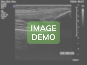
The infrapatellar fat pad shows the normal ultrasound features of mature adipose tissue.
Findings: Tessuto adiposo ipereoico omogeneo con normale morfologia triangolare. Contorni regolari e ben definiti. Assenza di ispessimenti, bande fibrotiche o aree di fibrosi. Non si evidenziano raccolte fluide o alterazioni strutturali. Vascolarizzazione fisiologica al Doppler senza segni di iperemia o neoangiogenesi. Rapporti normali con il legamento rotuleo e le strutture circostanti.
🔄
🔄
🔄
Zone not present
ℹ️ Quadriceps tendon
In this case, the quadriceps tendon shows normal ultrasound features typical of a healthy adult.
Findings: Well-defined parallel fibrillar echotexture. Homogeneous ultrasound pattern without areas of thickening or hypoechogenicity. Regular myotendinous junction. Clear and well-defined insertion at the base of the patella. No intratendinous calcifications or vascular alterations on Doppler.
🔍
Zone not present
ℹ️ Patellar ligament
The patellar ligament shows completely normal morphology and echotexture for the patient’s age.
Findings: Length of 48 mm with a uniform thickness of 4 mm. Straight course from the patellar apex to the anterior tibial tuberosity. Normal tendinous echotexture with clearly visible parallel fibers. No areas of degeneration, calcification, or discontinuity. Regular bony insertions without signs of enthesopathy. Normal vascularization on Doppler.
🔍
🔍
🔍
VS
Anatomical Zone
VS