
Clinical Cases Comparison
GN-ANT-M-A1016 - Moderate
VS
GN-ANT-G-A1013 - Severe

Classification
Joint:
Knee
View:
Anterior
Severity:
Moderate
Patient
Age:
86 years
Sex:
Female
Condition:
Acquired hemophilia A (autoimmune, inhibitor positive)
Annotations

Classification
Joint:
Knee
View:
Anterior
Severity:
Severe
Patient
Age:
38 years
Sex:
Male
Condition:
Hemophilia A severe (FVIII <1%), chronic hemophilic arthropathy
Annotations

History
GN-ANT-M-A1016
Elderly female patient diagnosed with acquired hemophilia A at age 82 following spontaneous extensive soft tissue hematomas and prolonged aPTT discovered during preoperative evaluation for cataract surgery. Inhibitor titer measured at 18 Bethesda Units. No previous personal or family history of bleeding disorders. Has experienced 4 episodes of spontaneous right knee hemarthrosis over the past 30 months despite immunosuppressive therapy (corticosteroids and cyclophosphamide) and bypassing agent coverage. Joint symptoms complicate pre-existing age-related osteoarthritis. Reports moderate pain (VAS 5-6/10) with daily activities. Uses walker for ambulation. Living independently with home care assistance three times weekly.
GN-ANT-G-A1013
Patient with severe hemophilia A diagnosed in infancy with history of poor adherence to prophylaxis during adolescence and early adulthood. Right knee has been the primary target joint since age 10, with documented hemarthrosis frequency of 12-15 episodes annually during teenage years. Despite initiation of prophylaxis at age 22, continued breakthrough bleeding due to high-demand physical job (construction worker). Has undergone two arthrocentesis procedures for tense hemarthrosis evacuation. Reports constant chronic pain (VAS 7-8/10) requiring daily opioid analgesics. Significant functional impairment with inability to climb stairs, prolonged standing, or walking distances >100 meters. Currently on disability leave from work.
Physical Examination
GN-ANT-M-A1016
Moderate knee swelling with palpable effusion. Age-related muscle atrophy of lower extremities with quadriceps showing additional wasting from hemophilic involvement. Mild valgus knee deformity. Range of motion limited by combination of hemarthrosis sequelae and degenerative changes: lacks 15° extension, achieves 115° flexion with discomfort. Crepitus present throughout range. Joint line tenderness medially and anteriorly. Gait significantly impaired requiring walker support with slow, cautious pattern. Difficulty with sit-to-stand transfers.
GN-ANT-G-A1013
Severe knee deformity with fixed flexion contracture at 25°. Marked muscle wasting of entire thigh with quadriceps atrophy of 5cm compared to left leg. Palpable bony enlargement and marked synovial thickening throughout joint. Severe limitation of range of motion: fixed flexion deformity prevents full extension, maximum flexion limited to 85°. Joint instability evident with valgus deformity. Ambulation requires cane support with pronounced limp. Severe tenderness throughout anterior knee structures. Quadriceps lag sign positive with inability to perform straight leg raise against gravity.
Ultrasound Findings
GN-ANT-M-A1016
- Hoffa’s fat pad: moderate alterations combining hemophilic and degenerative changes
GN-ANT-G-A1013
- Quadriceps tendon: severe degenerative changes with extensive structural damage
Anatomical Zones
ℹ️ Hoffa’s fat pad
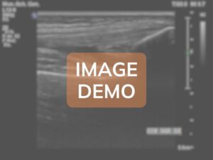
The adipose pad demonstrates moderate chronic alterations representing a complex interplay between acquired hemophilic arthropathy and age-related degenerative changes, with predominant hemosiderin deposition and fibrotic transformation.
Findings: The Hoffa's fat pad shows moderate pathological alterations with partial preservation of adipose tissue architecture. Approximately 50-60% of the fat pad volume maintains recognizable adipose echostructure (hyperechoic), while the remaining 40-50% demonstrates fibrotic transformation. The overall thickness measures 9.5mm, representing moderate enlargement compared to age-matched controls (expected 6-7mm in elderly patients). Multiple hypoechoic areas are distributed throughout the fat pad, with the largest measuring 12x7mm in the superior central portion, representing zones of fibrotic tissue replacement and hemosiderin deposition from recurrent hemorrhagic episodes. Several discrete hyperechoic foci without posterior shadowing are scattered within the fat pad, consistent with hemosiderin deposits rather than calcifications, appearing as 1-2mm punctate echogenic spots. Approximately 5-6 fibrous bands (thickness 1.5-2mm each) traverse the fat pad in various orientations, most prominently in the superior and lateral aspects, creating a reticular pattern that disrupts the normal homogeneous adipose appearance. These bands likely represent organized fibrous septa from previous hemorrhagic organization. The anterior border adjacent to the patellar ligament shows irregular interface with loss of the normal smooth plane, suggesting adhesion formation. The posterior border demonstrates moderate thickening and irregularity where it interfaces with the synovium. A small loculated fluid collection (5x3mm) is present in the lateral recess of the fat pad, possibly representing a small hemorrhagic cyst or loculated effusion. Color Doppler examination reveals moderate increase in vascularity with 4-5 small vessels (0.5-0.8mm diameter) penetrating into the fibrotic areas, indicating ongoing inflammatory activity despite the patient's age. The vascular pattern is less chaotic compared to younger patients with hemophilic arthropathy, possibly reflecting the slower disease tempo in acquired hemophilia. The fat pad demonstrates moderately reduced compressibility on probe pressure compared to expected in elderly patients, indicating significant fibrotic component. Dynamic examination during knee flexion reveals impaired mobility of the fat pad with restricted anterior displacement, contributing to anterior knee pain and impingement symptoms. The degree of fibrosis appears disproportionate for only 30 months of disease duration, suggesting that pre-existing age-related fat pad changes (fibrofatty involution commonly seen in elderly patients) may have been accelerated and exacerbated by the superimposed hemorrhagic episodes. Areas of the fat pad adjacent to the patellar ligament and tibial plateau show increased echogenicity and thickening, consistent with chronic mechanical stress and inflammation. No discrete calcifications or ossifications are identified, which is typical for the relatively short disease duration despite patient's age.
🔍

🔍
🔍

🔍
Zone not present
Zone not present
ℹ️ Quadriceps tendon
The quadriceps tendon demonstrates severe chronic degenerative changes with extensive structural destruction secondary to decades of recurrent hemorrhagic episodes, representing end-stage tendinopathic changes.
Findings: The quadriceps tendon demonstrates severe pathological changes throughout its entire length from the myotendinous junction to patellar insertion. Marked diffuse thickening measures 11.5mm in the central portion, representing more than 70% increase from normal values. The normal homogeneous parallel fibrillar echostructure is completely obliterated, replaced by a severely heterogeneous appearance with no recognizable organized tissue pattern. Extensive areas of mucoid degeneration appear as large confluent hypoechoic zones (largest measuring 15x8mm) occupying approximately 40% of the cross-sectional area, indicating advanced tendon necrosis. Multiple large intratendinous calcifications are present throughout the tendon substance: at least 8-10 distinct calcific deposits ranging from 3mm to 12mm in diameter, with the largest forming a continuous calcified plaque in the distal third. These calcifications demonstrate dense hyperechoic appearance with prominent posterior acoustic shadowing. Severe enthesopathy is evident at the patellar insertion with marked irregularity of the patellar base, multiple erosions (largest 8x5mm), and extensive reactive bone formation creating irregular osteophytic spurs. The superior pole of the patella shows severe cortical disruption with loss of normal bone architecture. Evidence of partial-thickness tear is present in the superficial fibers of the mid-substance, with a hypoechoic cleft extending 8mm in length and 4mm in depth, representing approximately 35% cross-sectional involvement. The deep fibers show severe disorganization but maintain some continuity. The myotendinous junction demonstrates abnormal thickening and irregularity with extension of pathological changes into the distal muscle belly. Extensive intratendinous fibrosis is evident as hyperechoic bands and nodules interspersed with the degenerative areas. Color and Power Doppler reveal marked pathological neovascularization with chaotic vascular pattern throughout the tendon, particularly concentrated around the areas of mucoid degeneration and at the enthesis. Multiple large feeding vessels (>1mm diameter) penetrate the tendon from both anterior and posterior surfaces, indicating active chronic inflammatory process. Peritendinous changes include marked thickening of the paratenon (4mm) with loss of normal gliding planes and extensive adhesions to surrounding tissues, particularly to the suprapatellar fat and prefemoral fat pad. Dynamic examination during attempted knee extension demonstrates severely impaired tendon excursion with rigid, non-elastic behavior and lack of normal lengthening-shortening pattern. The tendon appears functionally compromised with evidence of mechanical insufficiency contributing to the extensor lag observed clinically. Measurement of the anterior-posterior thickness shows asymmetric thickening with the anterior portion measuring 13mm and posterior portion 9mm, indicating heterogeneous degenerative process. Multiple focal areas of tendon discontinuity suggest previous partial rupture episodes with attempted healing through disorganized scar tissue formation.

🔍
🔍
VS
Anatomical Zone
VS