
Clinical Cases Comparison
CV-ANT-G-A1099 - Severe
VS
CV-ANT-L-A1097 - Mild

Classification
Joint:
Ankle
View:
Anterior
Severity:
Severe
Patient
Age:
46 years
Sex:
Male
Condition:
Hemophilia A severe (FVIII <1%), end-stage hemophilic arthropathy
Annotations

Classification
Joint:
Ankle
View:
Anterior
Severity:
Mild
Patient
Age:
62 years
Sex:
Male
Condition:
Hemophilia B moderate (FIX 3%)
Annotations
History
CV-ANT-G-A1099
Patient with severe hemophilia A diagnosed at 18 months following intracranial hemorrhage. Target joint development in right ankle began at age 7, with estimated 200+ hemarthrosis episodes throughout childhood and adolescence. Received on-demand treatment with cryoprecipitate until age 12, then plasma-derived concentrates. Prophylaxis initiated at age 18 but with poor adherence during young adulthood (age 18-28) due to psychosocial challenges. Compliance improved after age 30, but extensive joint damage already established. Reports severe chronic pain (VAS 8-9/10) requiring daily opioid analgesics. Previous interventions: radiosynovectomy at age 25, arthroscopic synovectomy at age 32 (temporary improvement). Currently on long-term disability, unable to work. Experiences 4-5 breakthrough bleeding episodes annually despite prophylaxis (FVIII 3000 IU three times weekly). Comorbidities include chronic pain syndrome, depression, hepatitis C (cured 2019), and hypertension.
CV-ANT-L-A1097
Patient diagnosed with moderate hemophilia B at age 8 following excessive bleeding after minor trauma. Received on-demand treatment throughout childhood and adolescence due to limited factor availability in the 1970s-80s. Started prophylaxis at age 45 after multiple joint bleeds. Right ankle has experienced 3 documented hemarthrosis episodes over past 5 years, all following minor trauma (twisting injury, stepping off curb). Most recent episode 8 months ago, resolved with factor replacement and RICE protocol within one week. Reports occasional mild ankle stiffness in the morning (resolves within 15-20 minutes) and mild discomfort after prolonged standing or walking >1 hour. Currently on prophylaxis with recombinant FIX 1500 IU twice weekly.
Physical Examination
CV-ANT-G-A1099
Severe ankle deformity with marked varus hindfoot alignment (15° varus). Massive swelling with tense effusion and prominent circumferential synovial thickening. Significant muscle atrophy of entire lower leg with calf circumference 6cm smaller than contralateral (25cm vs 31cm). Skin changes include hyperpigmentation and thinning. Fixed equinus contracture with ankle resting in 25° plantarflexion, unable to achieve neutral position. Range of motion severely restricted: complete loss of dorsiflexion (0°), plantarflexion limited to 25° from resting position (total arc only 25°). Rigid end-feel suggesting bony ankylosis and severe capsular contracture. Joint instability with medial-lateral laxity and positive anterior drawer. Severe tenderness throughout. Gait severely antalgic requiring bilateral forearm crutches. Cannot perform single-leg stance or any heel raises. Moderate pedal edema (2+) extending to mid-calf.
CV-ANT-L-A1097
Ankle inspection shows no obvious swelling or deformity. Minimal effusion detectable only with experienced palpation techniques. No erythema or warmth. Slight tenderness on deep palpation of anterior joint line. Range of motion mildly reduced: dorsiflexion 15° (normal 20°), plantarflexion 45° (normal 50°), with mild sense of anterior tightness at end-range dorsiflexion. Muscle strength intact with 5/5 throughout. Gait normal without limp. Able to perform single-leg heel raises ×15 repetitions (slight fatigue compared to expected ×20). Single-leg stance >25 seconds, slightly reduced from optimal. No ligamentous instability on stress testing.
Ultrasound Findings
CV-ANT-G-A1099
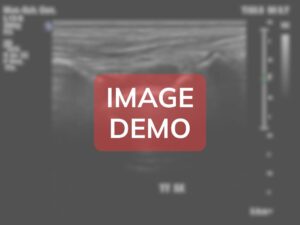
Ultrasound examination in anterior view reveals severe end-stage pathological changes:
- Anterior joint capsule: severe thickening (>8mm) with calcifications
- Anterior recess: massive synovial proliferation (>10mm) with complex organized effusion
- Talar dome: severe cartilage loss with near-complete destruction and exposed bone
- Distal tibial surface: extensive erosions, large osteophytes, subchondral cysts
- Marked synovial hyperemia with chaotic neovascularization
- Bony surfaces: extensive erosions, irregular contours, pseudocysts
- Large complex effusion with multiple loose bodies and debris
- Anterior soft tissue severe thickening and fibrosis
- Joint space collapse with areas of pseudoankylosis
CV-ANT-L-A1097
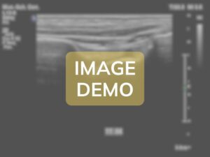
Ultrasound examination in anterior view reveals early changes:
- Anterior joint capsule: mild thickening (2.5mm)
- Anterior recess: minimal synovial proliferation, trace effusion
- Talar dome: early cartilage surface irregularities
- Distal tibial surface: minimal changes
- Mild synovial thickening with minimal hyperemia
- Bony surfaces: minimal cortical irregularity
- No significant osteophytes or erosions
- Extensor tendons: normal appearance
Anatomical Zones
🔄 Tibiotalar joint anterior recess
The anterior recess demonstrates severe end-stage degenerative changes consistent with long-standing severe hemophilic arthropathy, showing massive synovial proliferation, near-complete articular cartilage destruction, extensive bony erosions, and structural disorganization representing decades of recurrent hemorrhagic insults.
Findings: The anterior recess displays catastrophic pathological alterations representing end-stage disease. The synovial membrane demonstrates massive proliferation measuring 11.5mm (normal 80% loss). Multiple large full-thickness defects (10+ areas ranging 5-15mm) expose severely irregular, eroded bone surfaces. The largest defect (15x8mm) creates deep excavation into subchondral bone. The talar subchondral bone shows catastrophic changes with multiple large erosions (8-12mm depth) creating "moth-eaten" appearance. Numerous subchondral pseudocysts (5-10mm diameter) represent bone necrosis and cystic degeneration. The distal tibial surface demonstrates similarly severe destruction with multiple erosions (5-12mm depth), near-complete cartilage loss (80% loss) with areas of apparent bone-on-bone contact suggesting pseudoankylosis. The anterior capsule demonstrates severe fibrotic thickening (8.5mm) with 15-20 calcific deposits (1-4mm) scattered throughout. Color Doppler reveals marked neovascularization with >15-20 vessels (0.5-2mm) creating dense chaotic network (grade 3-4 vascularity). Power Doppler shows confluent synovial blush. Spectral Doppler demonstrates low-resistance flow (RI 0.45-0.55) with elevated velocities (15-25 cm/sec) indicating active inflammation. Peri-articular soft tissues show severe thickening (8-10mm subcutaneous tissue) with obliterated tissue planes and widespread tenosynovitis in multiple extensor tendon sheaths.

🔄

🔄

🔄
🔄 Tibiotalar joint anterior recess
The anterior recess demonstrates early pathological changes consistent with mild hemophilic arthropathy, showing initial synovial proliferation and subtle articular surface alterations from recurrent low-grade hemorrhagic insults.
Findings: The anterior recess of the tibiotalar joint shows mild pathological alterations indicating early hemophilic involvement. The synovial membrane demonstrates mild thickening measuring 2.8mm (normal <1mm), appearing as a hypoechoic band lining the anterior capsule. The synovial echostructure is slightly heterogeneous rather than uniformly thin, suggesting early synovial proliferation. A small amount of anechoic to minimally hypoechoic fluid (approximately 2mm in anterior-posterior dimension) is visible within the recess, representing trace joint effusion. This fluid contains few internal echoes, suggesting relatively recent or chronic effusion without acute hemorrhagic components. The talar dome cartilage shows early surface irregularities with loss of the perfectly smooth contour seen in normal joints. The cartilage thickness appears relatively preserved (1.3-1.5mm) but the superficial layer demonstrates focal areas of slight irregularity, particularly in the central weight-bearing zone, measuring approximately 4x2mm. These irregularities appear as subtle undulations of the cartilage-fluid interface rather than frank erosions. The underlying subchondral bone of the talus maintains its hyperechoic cortical line, though very close inspection reveals minimal irregularity (1-2 small areas of 0.5mm cortical discontinuity) suggesting early erosive changes. The distal tibial articular surface appears relatively well-preserved with only minimal irregularity of the anterior lip, where a small area (2x1mm) shows slight cortical irregularity without frank osteophyte formation. The joint space width remains within normal limits (3-4mm) with preserved parallel alignment of articular surfaces, though slight asymmetry is noted with marginally wider spacing medially (3.8mm) versus laterally (3.2mm). Two small hyperechoic foci (1mm each) are visible within the anterior recess, possibly representing early hemosiderin deposits or tiny debris particles from previous hemorrhagic episodes. Color Doppler examination reveals mild increase in synovial vascularity with 2-3 small vessels (0.3-0.5mm diameter) penetrating the synovial membrane, indicating low-grade inflammatory activity. The vascular pattern is organized and sparse rather than the chaotic hypervascular pattern seen in more severe disease. Power Doppler confirms the presence of flow within the thickened synovium, with signal intensity grade 1 (minimal vascularity). The anterior joint capsule shows mild thickening (2.5mm versus expected <2mm) with slightly irregular fibrous architecture, though overall continuity is maintained. The anterior tibial lip demonstrates minimal irregular thickening without definitive osteophyte formation - this represents early reactive change rather than established degenerative alterations. Dynamic examination during passive dorsiflexion shows the anterior recess opening normally, though the synovial tissue appears slightly less mobile with subtle restriction of normal fold patterns. During plantarflexion, the recess collapses but maintains slightly more residual fluid than expected in a normal joint. No impingement or mechanical block is demonstrated during range of motion. The subcutaneous tissues anterior to the capsule appear normal without edema or inflammatory changes. The extensor retinaculum maintains normal thickness and echostructure. Overall, these findings represent early-stage hemophilic arthropathy with predominant synovial and superficial cartilage involvement, without established destructive changes to deeper structures.

🔄
🔄
VS
Anatomical Zone
VS